PhD opportunity [February 2024 start] on "Accurate automated quantification of spine evolution — it’s about time!"
Project overview:
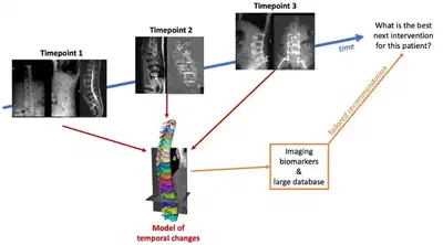
- Title: Accurate automated quantification of spine evolution — it’s about time!
- First supervisor: Marc Modat
- Second supervisor: Tom Vercauteren
- Clinical Supervisor: Amanda Isaac
- Start date: February 2024
Aim of the PhD Project
- Complement radiological expertise with automated analysis of longitudinal spine changes
- Development of longitudinal registration algorithms to align spine images from different imaging modalities
- Development of processing tools robust to the presence of metal artefact and various fields of view
- Extraction of imaging biomarkers of spine degeneration.
Project summary
Back pain presents because of a wide range of conditions in the spine and is often multifactorial. Spine appearances also change after surgery, and postoperative changes depend on the specific interventions offered to patients and human factors such as healing and mechanical adaptations, which are also unique to each patient. When reviewing medical images for diagnostic, monitoring or prognosis purpose, radiologists are required to evaluate multiple structures, including bone, muscles, and nerves, as well as the surrounding soft tissues and any instrumentation used in surgery. They must use their expertise to assess how each structure has evolved over time visually. Such readings are, therefore, both time-consuming and require dedicated expertise, which is limited to large regional spine centres throughout the UK.
This project aims to develop a tool to characterise and quantify the changes occurring in an individual’s spine over time. It will take advantage of the many medical images, mostly computer tomography scans and magnetic resonance images, that are acquired per patient as they go through several surgical procedures. Imaging biomarkers will then be extracted and, used in conjunction with other information about the patient, will assist clinical teams in providing improved care to their patients.
A large database of images from patients who underwent surgery at Guys’ and St Thomas’ Trust hospital between 2012 and 2022 will be used to develop, train, and validate machine learning models capable of tracking the changes in a patient’s spine. These models will be required to perform well in the presence of metal artefact induced by implants. They will also need to cope with the different imaging modalities and the various fields of view of the acquired images, where some cover the whole spine and others only a subset. A dedicated geometrical model must also be tailored to the geometry of the spine, including articulated rigid structures, the vertebrae, surrounded by soft tissues such as discs, muscles, fat or nerves.
Additionally, a better understanding of surgery-related changes will provide valuable information to guide and assess interventions. Faster and more objective demonstration of changes in spine depicted on imaging may also lead to more efficient diagnostic pathways, which are patient-specific at a more value-based, time and cost-efficient model to an overburdened limited diagnostic national service.
The candidate should have a background in one of the following: biomedical engineering, applied mathematics/physics or computer science. The candidate should also have a keen interest in medical image processing, advanced machine learning and modelling.
More information about the PhD project and how to apply on the website of the EPSRC Centre for Doctoral Training in Smart Medical Imaging.
Tom Vercauteren
Professor of Interventional Image Computing
Tom’s research interests include machine learning and computer assisted interventions