PhD opportunity [June 2026 start] on "Anatomy localisation in X-ray fluoroscopy videos for mechanical thrombectomy" in collaboration with Telos Health
Applications are invited for a fully funded 4 years PhD EPSRC CDT PSI studentship (including tuition fees, annual stipend and consumables) starting in June 2026.
Award details:
- Focus: Anatomy localisation in X-ray fluoroscopy videos for mechanical thrombectomy
- First supervisor: Tom Vercauteren
- Second supervisor: Thomas Booth
- Industry supervisor: Konrad Leibrandt, Telos Health
- Funding type: 4-year fully-funded EPSRC CDT PSI studentship including a stipend, tuition fees, research training and support grant (RTSG), and a travel and conference allowance.
- Start date: June 2026
Project description
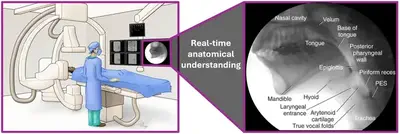
This project focuses on the development of a learning-based system capable of localising anatomy in X-Ray fluoroscopy video streams acquired during mechanical thrombectomy (MT). Mechanical thrombectomy are emergency procedures for acute ischaemic stroke. An endovascular catheter is navigated from the groin up to the brain under real-time X-ray fluoroscopy guidance. MT is challenging to perform in part due to the complexity of fluoroscopy image interpretation. Computer vision approaches have shown promising capabilities in surgical scene understanding across several minimally invasive surgical specialties but their development in interventional neuroradiology remains at its infancy. The PhD candidate will develop novel methods to automatically identify anatomical structures in fluoroscopy videos with the ambition of providing a better spatial understanding to the interventional neuroradiologist (INR). This project will require close collaboration with expert INRs to build annotated fluoroscopy databases and validate the performance of the proposed solutions.
Application Process
Tom Vercauteren
Professor of Interventional Image Computing
Tom’s research interests include machine learning and computer assisted interventions