PhD opportunity [June 2022 start] on "Computational approaches for quantitative fluorescence-guided neurosurgery"
Applications are invited for the fully funded 4 years full-time PhD studentship (including home tuition fees, annual stipend and consumables) starting on 1st June 2022.
Award details:
- Focus: Translational research on hyperspectral-based quantitative fluorescence imaging linked with a neurosurgery clinical study
- Primary supervisor: Tom Vercauteren
- Secondary supervisor: Jonathan Shapey
- Industry collaborator: Hypervision Surgical
- Funding type: Tuition fee, stipend (Home Fee status only).
- Application closing date: 11 April 2022
- Start date: June 2022
Aim of the project
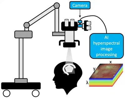
This project aims at enabling wide-field and real-time quantitative assessment of tumour-specific fluorescence by designing novel deep-learning-based computational algorithms. The project will leverage a compact hyperspectral imaging (HSI) system developed by Hypervision Surgical Ltd initially designed for contrast-free imaging.
The success of brain tumour surgery is largely dependent on how much of the tumour can be safely removed during surgery. Using HSI as an advanced optical imaging technique, early research results with slow benchtop HSI systems have shown that it is possible to extract quantitative information about fluorophore concentration and hence about tumour burden.
The primary hypothesis being tested in this project is that HSI-based quantitative fluorescence can be done in real-time with a device suitable for integration into the surgical workflow thanks to learning-based computational approaches.
Project details
Background. Brain tumours pose a significant public health burden, with 70,500 patients diagnosed with brain tumours in the UK each year. The success of brain tumour surgery is largely dependent on two key factors: how early the tumour is detected, and how much of the tumour can be safely removed during surgery. However, determining brain tumour from healthy brain tissue can be exceedingly difficult during surgery. To significantly improve patient outcomes, neurosurgeons need a way to reliably identify early-stage low-grade gliomas during surgery.
Recent developments in smart camera systems such as hyperspectral imaging can enhance the surgeon’s vision. Hyperspectral imaging could help neurosurgeons to detect the low levels of fluorescence generated by early-stage, low-grade gliomas and guide the removal of the tumour to improve patient outcomes. However, the data generated by hyperspectral imaging cameras is complex and requires advanced computational processing to be useful for surgical guidance.
Addressing these challenges and limitations of the previous generation of smart cameras, this project aims to develop real-time quantitative fluorescence HSI (qFHSI) algorithms that will eventually deliver a step-change in the treatment of LGG. It will involve collaborations with neurosurgeons, hardware and software engineers from KCL and the project’s industrial partner, Hypervision Surgical Ltd.
Year 1: The student will receive appropriate skills training to enable them to conduct the research project. Part of this aim will be achieved by performing a literature review on the existing quantitative fluorescence approaches. Building on previous work in the group on developing a benchtop HSI-based system for quantitative fluorescence (Xie 2017), the student will get familiar with the particularities of real-time intraoperative HSI (Ebner 2021) and adapt the existing state-of-the-art algorithms to this platform.
Year 2: While the main technical deliverable of Year 1 is a first system for real-time quantitative fluorescence using a snapshot imaging device, it would have been developed by adapting computational approaches initially designed for high-resolution spectral data acquired across a wide wavelength range. While of interest, this initial adaptation is nonetheless expected to lead to suboptimal results. In Year 2, advanced data-driven computational biophotonics approaches will allow the design of a tissue-optics model that can compensate for scattering and absorption at wavelengths not directly captured by the captured fluorescence bands. Physics-informed deep learning will be combined with experimental measurements and Monte Carlo simulations to achieve accurate and robust extrapolation. To support this goal, the student will spend a 3-month placement at Hypervision Surgical where they will get support on the physics of acquisition of the imaging system. In addition, the student will get hands-on experience with the development of algorithms and imaging systems as part of a regulated medical device development.
Year 3: Further refinement of the quantitative fluorescence model will be made in Year 3. In particular, the computational model will be extended to incorporate tissue autofluorescence and account for spatio-spectral subsampling. Integration and optimisation of the algorithm will be key to achieve real-time quantitative fluorescence measurement. Validation of the system will be performed in bespoke phantoms to assess the limitations of the developed system.
Year 4: On the technical side, the final year will focus on consolidating the previous developments and addressing the limitations identified in Year 3 whenever feasible in the time frame of the PhD. User-centred validation will be performed by designing bespoke evaluation protocols implemented in a realistic surgical environment. Finally, time is set aside for contingency planning and for the write-up of the thesis. This will ensure the student finishes their PhD within 4 years.
Further information
Informal email enquiries from interested students to the supervisor are encouraged (contact details below).
Prof. Tom Vercauteren: [email protected]
Award value
The studentship is fully funded for 4 years. This includes home tuition fees, stipend and generous project consumables.
Stipend: Students will receive a tax-free stipend at the UKRI rate of ca £20,109 per year as a living allowance.
Research Training Support Grant (RTSG): A generous project allowance will be provided for research consumables and for attending UK and international conferences. Eligibility criteria
Prospective candidates should have a 1st or 2:1 M-level qualification in Biomedical Engineering, Physics, Engineering, Computer Science, Mathematics, or a related programme.
Preference will be given to candidates with a background conducive to multidisciplinary research and preferably programming skills.
Candidates who meet the eligibility requirements for Home Fee status will be eligible to apply for this project. Home students will be eligible for a full UKRI award, including fees and stipend, if they satisfy the UKRI criteria below, including residency requirements. To be classed as a Home student, candidates must meet the following criteria:
- be a UK National (meeting residency requirements), or
- have settled status, or
- have pre-settled status (meeting residency requirements), or
- have indefinite leave to remain or enter.
We welcome eligible applicants from any personal background, who are pleased to join diverse and friendly research groups.
Applicable level of study: Postgraduate research
Application process
Please submit an application for the Biomedical Engineering and Imaging Science Research MPhil/PhD (Full-time) programme using the King’s Apply system. Please include the following with your application:
- A PDF copy of your CV should be uploaded to the Employment History section.
- A 500-word personal statement outlining your motivation for undertaking postgraduate research should be uploaded to the Supporting statement section.
- Funding information: Please choose Option 5 “I am applying for a funding award or scholarship administered by King’s College London” and under “Award Scheme Code or Name” enter MRC_TV. Failing to include this code might result in you not being considered for this funding
Tom Vercauteren
Professor of Interventional Image Computing
Tom’s research interests include machine learning and computer assisted interventions
Jonathan Shapey
Clinical Academic and Consultant Neurosurgeon
Jonathan’s academic interest focuses on the application of medical technology and artificial intelligence to neurosurgery.
Michael Ebner
CEO and co-founder, Hypervision Surgical
Michael is an entrepreneur and a scientist focusing on the development of computational hyperspectral imaging for real-time surgical guidance to improve surgical precision and patient safety.