PhD opportunity on "Artificial intelligence-driven management of brain tumours"
Post overview:
- Title: Artificial intelligence-driven management of brain tumours
- Project ID: BE-MI2023_15
- First supervisor: Jonathan Shapey
- Second supervisor: Tom Vercauteren
- Start date: October 2023
- Application deadline: Wednesday 9 November 2022, 1:00 PM (GMT)

Project description
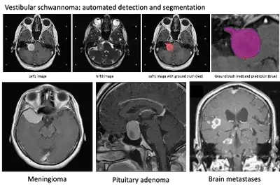
Approximately 25,000 patients are diagnosed with a brain tumour every year in the UK. Meningiomas and pituitary adenomas are the first and third most common primary tumour, accounting for over 50% of all primary brain tumours. Brain metastases affect up to 40% of patients with extracranial primary cancer.
Patients with brain tumours require individualized patient management. The automated detection and segmentation of brain tumours could help personalise and standardise patient management and significantly improve clinical workflow.
We have previously developed a fully-automated AI framework to segment a vestibular schwannoma (a type of brain tumour) from MRI achieving state-of-the-art results. This project aims to develop deep learning models to: 1) detect and automatically segment various non-glial brain tumours (meningioma, pituitary adenoma, brain metastases) using MRI; and 2) develop compositive clinical and imaging biomarkers to predict tumour growth and behaviour (pituitary adenoma, meningioma).
In Year 1 the student will learn modern image-registration and domain-adaptation methods to curate a complete dataset of registered magnetic resonance images using the patient’s available sequences. Year 2 will include investigation of radiomic data extraction and automated tumour classification methods using machine learning. In Year 3, the student will develop data-driven deep learning frameworks combining longitudinal clinical and imaging data to create radiomic biomarkers to predict tumour growth and behaviour. The workplan may be adjusted to accommodate the student’s background.
The student will receive specialised training in modern deep learning approaches and will acquire an in-depth understanding of how to apply these methods in medical imaging analysis.
One representative publication from each co-supervisor
- Shapey J, Wang G, Dorent R, Dimitriadis A, Li W, Paddick I, Kitchen N, Bisdas S, Saeed SR, Ourselin S, Bradford R, Vercauteren T. An artificial intelligence framework for automatic segmentation and volumetry of vestibular schwannoma from contrast-enhanced T1-weighted and high-resolution T2-weighted MRI. J Neurosurg 6:1-9 (2019) doi:10.3171/2019.9.JNS191949
- Dorent R, Booth T, Li W, Sudre CH, Kafiabadi S, Cardoso J, Ourselin S, Vercauteren T. Learning joint segmentation of tissues and brain lesions from task-specific hetero-modal domain-shifted datasets. Med Image Anal. 2021 Jan;67:101862. doi: 10.1016/j.media.2020.101862. Epub 2020 Oct 9. PMID: 33129151; PMCID: PMC7116853. doi:10.1016/j.media.2020.101862
Eligibility information
This project is offered as part of the MRC Doctoral Training Partnership (DTP) in Biomedical Sciences.
Home, EU and international students are all eligible to apply. The MRC DTP has funding available for up to 30% of its cohort for EU and international applicants. That is approximately 10 studentship places per year. All MRC DTP studentships are fully funded; including tuition fees, stipend and bench fee. If you are unsure if you meet the eligibility criteria, you can read the FAQ and contact the MRC DTP Admissions Team for clarification.
More information about the position here and how to apply here.