PhD opportunity [October 2026 start] on "Real-time intraoperative polarimetric imaging to detect nerves during brain tumour surgery"
Applications are invited for a fully funded 1+3 years MRes+PhD or 4 years PhD MRC DTP studentship (including tuition fees, annual stipend and consumables) starting in October 2026.
Award details:
- Focus: Real-time intraoperative polarimetric imaging to detect nerves during brain tumour surgery
- First supervisor: Jonathan Shapey
- Second supervisor: Tom Vercauteren
- Third supervisor: Graeme Stasiuk
- Funding type: 4-year fully-funded MRC DTP studentship including a stipend, tuition fees, research training and support grant (RTSG), and a travel and conference allowance.
- Application closing date: 28 October 2025
- Start date: October 2026

Project description
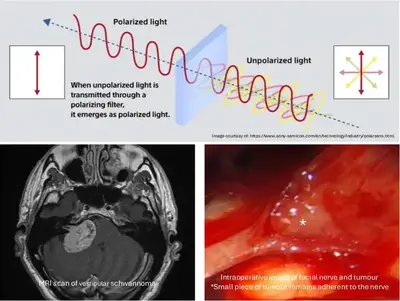
Vestibular schwannoma (VS) is the commonest pathology encountered in skull base neurosurgery. The goal of modern VS surgery is maximal tumour removal whilst preserving neurological function, but facial paralysis injury still occurs in 34% of patients because it is incredibly difficult for surgeons to visualise the facial nerve clearly during surgery.
Polarisation is a fundamental but under-exploited characteristic of light. Recent studies have led to new ways to identify nerves via their special polarization signature. Previous work has focussed on the characterisation of normal anatomy using devices that are challenging to integrate in the surgical workflow. This study aims to use a recently developed compact multi-polarisation-sensitive camera to achieve real-time intraoperative visualisation of nerve in the presence of tumour.
MRes 3-month research project: For the MRes project, students will compare the multi-polarisation-sensitive camera to a classical camera with a rotating polariser using standard polarised imaging scenes. Students will also attend the neurosurgical operating theatre to familiarise themselves with vestibular schwannoma pathology, surgery and the current limitations of standard microscopic visualisation.
Year 1: Characterisation of polarisation camera using phantom models Y1 will focus on setting up and characterising the imaging system based on the compact multi-polarisation-sensitive camera. A first pipeline for combining the multiple polarisation image responses will be designed. The student will research the physical and optical properties of nerve and tumour and develop realistic phantom models of these. The student will leverage the group’s extensive experience to fast-track developments.
Outcome: Imaging system for a detailed understanding of the optical polarisation of nerve and tumour
Year 2: Evaluation of optical polarisation of nerve and tumour in a murine model In Y2 the student will be trained in animal handling. Using an established murine model of sciatic nerve schwannoma, the student will fine-tune the combination algorithm and characterise performance in delineating nerve and tumour.
Outcome: Improved differentiation of nerve and schwannoma in vivo
Years 3 – 4: Development of a novel murine vestibular schwannoma model and evaluation of tissues using a polarisation camera In Y3–4, the student will collaborate with experts at the University of Manchester to develop a novel murine model of vestibular schwannoma. The student will perform stereotactic tumour implantation in the cerebellopontine angle and will then characterise nerve and tumour tissue in situ, mimicking vestibular schwannoma surgery.
Outcome: First demonstration of nerve and vestibular schwannoma differentiation in situ in a novel murine model.
Representative Publications from Supervisors
- MacCormac, O., Horgan, C. C., Waterhouse, D., Noonan, P., Janatka, M., Miles, R., … & Shapey, J. (2025). Hyperspectral abdominal laparoscopy with real-time quantitative tissue oxygenation imaging: a live porcine study. Frontiers in Medical Technology, 7, 1549245
- MacCormac, O., Noonan, P., Janatka, M., Horgan, C. C., Bahl, A., Qiu, J., … & Shapey, J. (2023). Lightfield hyperspectral imaging in neuro-oncology surgery: an IDEAL 0 and 1 study. Frontiers in Neuroscience, 17, 1239764
- Shapey, J., Xie, Y., Nabavi, E., Ebner, M., Saeed, S. R., Kitchen, N., … & Vercauteren, T. (2022). Optical properties of human brain and tumour tissue: an ex vivo study spanning the visible range to beyond the second near‐infrared window. Journal of biophotonics, 15(4), e202100072
- Li, P., Asad, M., Horgan, C., MacCormac, O., Shapey, J., & Vercauteren, T. (2023). Spatial gradient consistency for unsupervised learning of hyperspectral demosaicking: application to surgical imaging. International journal of computer assisted radiology and surgery, 18(6), 981-988
- Budd, C., Qiu, J., MacCormac, O., Huber, M., Mower, C., Janatka, M., … & Vercauteren, T. (2023, October). Deep reinforcement learning based system for intraoperative hyperspectral video autofocusing. In International Conference on Medical Image Computing and Computer-Assisted Intervention (pp. 658-667). Cham: Springer Nature Switzerland
- Ebner, M., Nabavi, E., Shapey, J., Xie, Y., Liebmann, F., Spirig, J. M., … & Vercauteren, T. (2021). Intraoperative hyperspectral label-free imaging: from system design to first-in-patient translation. Journal of Physics D: Applied Physics, 54(29), 294003