Closed Position

PhD opportunity [October 2024 start] on "Text promptable semantic segmentation of volumetric neuroimaging data"

Applications are invited for the fully funded 4 years full-time PhD studentship (including home tuition fees, annual stipend and consumables) starting on 1st June 2022.

Award details:

Text promptable semantic segmentation of volumetric neuroimaging data.

Project Overview

Semantic segmentation of brain structures from medical images, in particular Magnetic Resonance Imaging (MRI), plays an important role in many neuroimaging applications. Deep learning based segmentation algorithms are now achieving state-of-the-art segmentation results but currently require large amounts of annotated data under predefined segmentation protocols and data inclusion/exclusion criteria. The rigidity of such approaches forbids natural interactions by humans and thus limits the usefulness for non-routine questions.

Research Associate / Research Fellow in "Computational Hyperspectral Imaging"

Post overview:

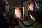
State-of-the-art fluorescence imaging system for neurosurgical guidance.

Job description

We are seeking an interventional image computing researcher to design and translate the next generation of AI-assisted hyperspectral imaging systems for surgical guidance using quantitative fluorescence. The postholder, based within the Department of Surgical & Interventional Engineering at King’s College London, will play a key role in NeuroPPEye a collaborative project with King’s College Hospital and work closely with the project’s industrial collaborator Hypervision Surgical, a King’s spin-out company. A clinical neurosurgery study underpins this collaboration. The successful candidate will work on the resulting neurosurgical data as well as controlled phantom data. They will also have the opportunity to provide insight on how to best acquire prospective data.

Research Associate / Research Fellow in "Trustworthy Artificial Intelligence for Surgical Imaging and Robotics"

Post overview:

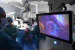
A view on a lumbar microdiscectomy surgery  (Source: [DVIDS Public Domain Archive](https://nara.getarchive.net/media/luis-contreras-a-contractor-orthopedic-technician-c6cb2a)).
A view on a lumbar microdiscectomy surgery (Source: DVIDS Public Domain Archive).

Job description

We are seeking a Post-doctoral Research Associate to develop novel trustworthy artificial intelligence (AI) algorithms able to extract actionable information from surgical imaging data.

[Job] Research Coordinator - King's College Hospital NHS Foundation Trust

We are seeking a motivated research nurse/coordinator to support our NeuroHSI and NeuroPPEye project.

CRN King’s Neurosurgery research coordinator

The post will be a Band 6 level Neurosurgery affiliated research nurse/coordinator to work within the neuroscience division at KCH. This is a full-time post, initially until end of August 2023 with a view to be extended by 6 - 12 months. The successful applicant will work across several neurosurgery sub-specialities with a particular focus on neuro-oncology and translational healthcare technology in neurosurgery. The applicate will work on research and clinical trials listed in the Department of Heath national portfolio, principally involving the development and evaluation of advanced smart camera technology for use during surgery. The post holder will work under the supervision of Mr Jonathan Shapey (Senior Clinical Lecturer and Consultant Neurosurgeon), Professor Keyoumars Ashkan (Professor of Neurosurgery) and the management of Alexandra Rizos, Neuroscience Research Manager. Some experience in clinical research and knowledge of good clinical practice would be beneficial.

PhD opportunity [February 2024 start] on "Incorporating Expert-consistent Spatial Structure Relationships in Learning-based Brain Parcellation"

Project overview:

Human-AI trust can be defined as the belief that the AI system will satisfy a set of contracts of trust. This project will establish contracts of trust about the spatial relationships across brain structures.

Aim of the PhD Project

  • Develop trustworthy deep learning-based brain segmentation/parcellation.
  • Formalise trustworthiness as contracts on spatial relationships between labels that the algorithm must fulfil.
  • Establish mathematical/algorithmic frameworks to guarantee that the proposed segmentation/parcellation respect the contracts of trust.
  • Implement, validate, and disseminate the proposed algorithms using open-access datasets.

Project summary

Automated segmentation and labelling of brain structures from medical images, in particular Magnetic Resonance Imaging (MRI), plays an important role in many applications ranging from surgical planning to neuroscience studies. For example, in Deep Brain Stimulation (DBS) procedures used to treat some movement disorders, segmentation of the basal ganglia and structures such as the subthalamic nucleus (STN) can help with precise targeting of the neurostimulation electrodes being implanted in the patient’s brain. Going beyond segmentation of a few discrete structures, some applications required a full brain parcellation, i.e., a partition of the entire brain into a set of non-overlapping spatial regions of anatomical or functional significance. Brain parcellation have notably been used to automate the trajectory planning of multiple intracranial electrodes for epilepsy surgery or to support the assessment of brain atrophy patterns for dementia monitoring.

PhD opportunity on "Artificial intelligence-driven radiosurgery planning for brain metastases"

Project overview:

Automated detection and segmentation of brain metastases using MRI for radiosurgery planning.

Aim of the PhD Project

  • Implement learning-based registration to curate a spatially-normalised dataset of MR images previously used to deliver stereotactic radiosurgery to brain metastases
  • Develop data-driven deep learning frameworks to automatically detect and segment brain metastases while allowing for interactive corrections
  • Develop imaging biomarkers to predict tumour response and behaviour following treatment

Project summary

Approximately 25,000 patients are diagnosed with a brain tumour every year in the UK. Brain metastases affect up to 40% of patients with extracranial primary cancer. Furthermore, although there are presently no reliable data, metastatic brain tumours are thought to outnumber primary malignant brain tumours by at least 3:1. Patients with brain metastases require individualized patient management and may include surgery, stereotactic radiosurgery, fractionated radiotherapy and chemotherapy, either alone or in combination.

PhD opportunity [February 2024 start] on "Accurate automated quantification of spine evolution — it’s about time!"

Project overview:

Aiming at characterising and quantifying the changes occurring in an individual’s spine over time.

Aim of the PhD Project

  • Complement radiological expertise with automated analysis of longitudinal spine changes
  • Development of longitudinal registration algorithms to align spine images from different imaging modalities
  • Development of processing tools robust to the presence of metal artefact and various fields of view
  • Extraction of imaging biomarkers of spine degeneration.

Project summary

Back pain presents because of a wide range of conditions in the spine and is often multifactorial. Spine appearances also change after surgery, and postoperative changes depend on the specific interventions offered to patients and human factors such as healing and mechanical adaptations, which are also unique to each patient. When reviewing medical images for diagnostic, monitoring or prognosis purpose, radiologists are required to evaluate multiple structures, including bone, muscles, and nerves, as well as the surrounding soft tissues and any instrumentation used in surgery. They must use their expertise to assess how each structure has evolved over time visually. Such readings are, therefore, both time-consuming and require dedicated expertise, which is limited to large regional spine centres throughout the UK.

PhD opportunity [February 2024 start] on "Physically-informed learning-based beamforming for multi-transducer ultrasound imaging"

Project overview:

Multiple transducer delay-and-sum beamforming scheme (Peralta et al, 2019). It requires accurate transducer geometry to calculate the time-of-flight between each element and focal point, and apply proper time delays to each radio-frequency channel.

Aim of the PhD Project

  • Pursue sparse solutions to handle the channel count required to coherently operate multiple ultrasound transducer and design and implement machine learning strategies to avoid the sparsity-related artefacts in the images.
  • Develop advanced beamforming techniques using machine learning approaches informed by ultrasound physics to address the concern of the flexible geometry in a multi-transducer imaging system and achieve unprecedented image quality.
  • Explore the application of the techniques on healthy volunteers.

Project description

Medical Ultrasound (US) is a low-cost imaging method that is long-established and widely used for screening, diagnosis, therapy monitoring, and guidance of interventional procedures. However, the usefulness of conventional US systems is limited by physical constraints mainly imposed by the small size of the handheld probe that lead to low-resolution images with a restricted field of view and view-dependent artefacts.

PhD opportunity on "Artificial intelligence-driven management of brain tumours"

Post overview:

Exemplar cases and task for AI-driven management of brain tumours.

Project description

Approximately 25,000 patients are diagnosed with a brain tumour every year in the UK. Meningiomas and pituitary adenomas are the first and third most common primary tumour, accounting for over 50% of all primary brain tumours. Brain metastases affect up to 40% of patients with extracranial primary cancer.

Research Associate in "Biomedical Optics - Hyperspectral Imaging"

Post overview:

AI-assisted hyperspectral imaging systems for surgical guidance using quantitative fluorescence.

Job description

We are seeking a biomedical optics researcher to design and translate the next generation of hyperspectral imaging systems for surgical guidance using quantitative fluorescence. The postholder, based within the Department of Surgical & Interventional Engineering at King’s College London, will play a key role in a collaborative project with King’s College Hospital and work closely with the project’s industrial collaborator Hypervision Surgical, a recently founded King’s spin-out company. A clinical neurosurgery study has been set up to underpin this collaboration.