Posts
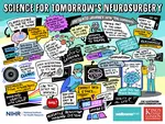
On 21st September we held our fourth ‘Science for Tomorrow’s Neurosurgery’ PPI group meeting online, with presentations from Oscar, Matt and Silvère. Presentations focused on an update from the NeuroHSI trial, with clear demonstration of improvements in resolution of the HSI images we are now able to acquire; this prompted real praise from our patient representatives, which is extremely reassuring for the trial going forward. We also took this opportunity to announce the completion of the first phase of NeuroPPEYE, in which we aim to use HSI to quantify tumour fluorescence beyond that which the human eye can see. Discussions were centered around the theme of “what is an acceptable number of participants for proof of concept studies,” generating very interesting points of view that ultimately concluded that there was no “hard number” from the patient perspective, as long as a thorough assessment of the technology had been carried out. This is extremely helpful in how we progress with the trials, particularly NeuroPPEYE, which will begin recruitment for its second phase shortly. Once again, the themes and discussions were summarized in picture format by our phenomenal illustrator, Jenny Leonard (see below) and we are already making plans for our next meeting in February 2024!
This video presents work lead by Martin Huber. Deep Homography Prediction for Endoscopic Camera Motion Imitation Learning investigates a fully self-supervised method for learning endoscopic camera motion from readily available datasets of laparoscopic interventions. The work addresses and tries to go beyond the common tool following assumption in endoscopic camera motion automation. This work will be presented at the 26th International Conference on Medical Image Computing and Computer Assisted Intervention (MICCAI 2023).
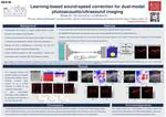
This video presents work lead by Mengjie Shi focusing on learning-based sound-speed correction for dual-modal photoacoustic/ultrasound imaging. This work will be presented at the 2023 IEEE International Ultrasonics Symposium (IUS).
You can read the preprint on arXiv: 2306.11034 and get the code from GitHub.
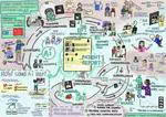
Recently, we organized a Public and Patient Involvement (PPI) group with Vestibular Schwannoma patients to understand their perspectives on an patient-centered automated report. Partnering with the British Acoustic Neuroma Association (BANA), we recruited participants by circulating a form within the BANA community through their social media platforms.
Post overview:
- Focus: Translational research on hyperspectral-based quantitative fluorescence imaging linked with a neurosurgery clinical study
- Line manager: Tom Vercauteren
- Clinical collaborator: King’s College Hospital
- Industry collaborator: Hypervision Surgical
- Salary: Grade 6, £41,386-£48,414 or Grade 7 £49,737-£55,306 per annum, including London Weighting Allowance


![PhD opportunity [October 2024 start] on "Text promptable semantic segmentation of volumetric neuroimaging data"](/post/2023-10-04-textpromptseg-phd/featured_hu_74717dac224f3504.webp)
